Post-traumatic Stress Disorder (PTSD) and Cerebellum | 17 Jan 2024
Why in News?
A recent study found that individuals with Post-traumatic Stress Disorder (PTSD) may experience significant decreases in both gray and white matter volume in their cerebellum.
- This could affect their cognitive functions and emotional responses, among other aspects.
What are the Findings of the Study?
- The study showed that PTSD is linked with considerable reductions in both gray and white matter volumes in the cerebellum.
- This reduction was particularly notable in specific subregions, including the posterior lobe, vermis, flocculonodular lobe and corpus medullare.
- The study also showed that cerebellar volume changes correlate with the intensity of the PTSD experience, offering a potential biomarker for assessing the condition’s severity.
- It challenges the traditional understanding of PTSD as solely a disorder of the brain’s emotion-processing centers.
- The cerebellum’s involvement suggests a more complex brain network disruption in PTSD, one that includes regions responsible for integrating cognitive and emotional responses.
- Study helps in understanding the pathophysiology of PTSD by pinpointing specific cerebellar regions affected by the disorder.
What is Post-Traumatic Stress Disorder (PTSD)?
- Post-traumatic Stress Disorder (PTSD), is a mental health condition that occurs after a person experiences or witnesses a traumatic event, such as war, violence, abuse, or natural disaster.
- People with PTSD may have intrusive memories, nightmares, flashbacks, avoidance and negative mood etc.
- These symptoms can interfere with their daily functioning and quality of life.
- PTSD can be treated with psychotherapy, medication, or both.
- PTSD is incredibly burdensome at both the individual and societal level, causing profound distress, functional impairment, and staggering treatment costs.
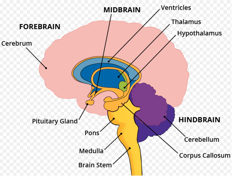
What is Cerebellum and Other Parts of the Brain?
- The brain comprises three primary components: the cerebrum, cerebellum, and brainstem.
- Cerebellum: The brain region traditionally associated with motor control, but now increasingly recognised for its role in higher cognitive and emotional functions.
- It is located at the back of the head, just below the cerebrum and behind the brain stem. Also called a “little brain” due to its similar but smaller structure than the cerebrum.
- Cerebrum: The largest part, consists of right and left hemispheres, playing a key role in higher functions like interpreting sensory information, speech, reasoning, emotions, learning, and precise movement control.
- Brainstem: Functioning as a relay centre connecting the cerebrum, cerebellum, and spinal cord. It oversees automatic processes such as breathing, heart rate, sleep-wake cycles, digestion, and various reflex actions like sneezing, coughing, vomiting, and swallowing.
- Hypothalamus: Situated below the thalamus and regulates functions including body temperature, hunger, thirst, fatigue, sleep, and circadian rhythms. It is also involved in the release of hormones by the pituitary gland.