Glow Scope | 13 Mar 2023
Why in News?
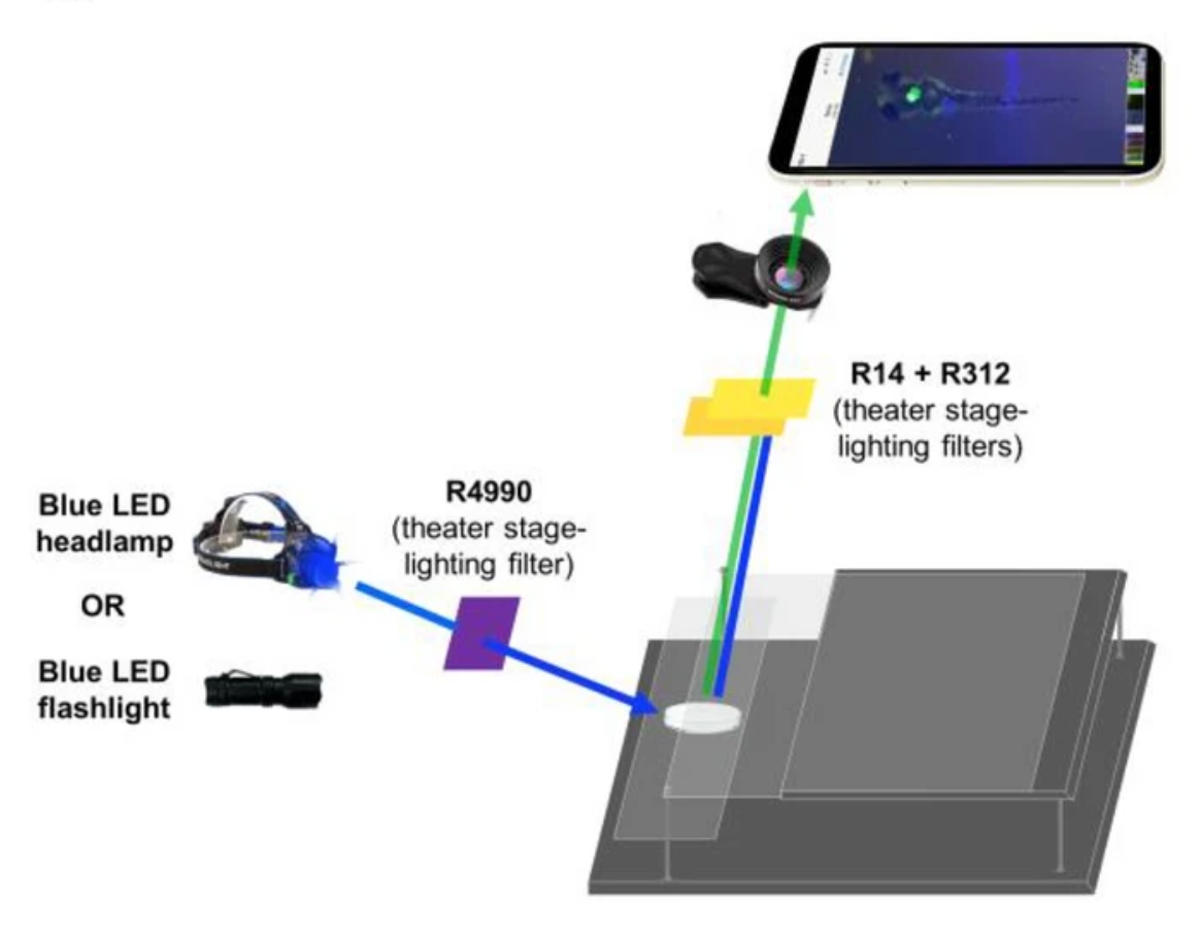
Researchers at Winona State University, Minnesota, have created a design for a Glow Scope, a Fluorescence Microscope.
- With this setup, they were able to image the creatures’ brain, spinal cord, heart, and head and jaw bones.
- They were able to zoom in and out using the smartphone camera and the clip-on lens.
What is Fluorescence Microscopy?
- About:
- An optical microscope views an object by studying how it absorbs, reflects or scatters visible light.
- A fluorescent microscope views an object by studying how it reemits light that it has absorbed, i.e., how it fluoresces. This is its basic principle.
- The object is illuminated with light of a specific wavelength. Particles in the object absorb this light and reemit it at a higher wavelength. These particles are called fluorophores; the object is infused with them before being placed under the microscope.
Is the Glowscope Accessible?
- Using a ‘glowscope’ still requires access to fluorophores, suitable biological samples, the know-how to combine the two, and some knowledge of physics to work out which LED flashlight to buy.
- The Foldscope was truly remarkable because all its required components were simple to understand.
- In 2014, a group of scientists at Stanford University released Foldscope, a handheld microscope made almost entirely out of paper, which takes 30 minutes to put together, and which could capture images of cells.
- However, the fact that a simple fluorescent microscope can be set up for a few thousand rupees means, instead of being entirely out of reach, researchers can prepare samples and take them to schools, where students can observe them.